Heart

Heart of dog in profile (left side):
1. Left ventricle. 2 Furrow with inter-ventricular coronary arteries. 3 right ventricle. 4 fat tissue. 5 pulmonary artery. 6 ligamentum arteriosum. 7 artery aorta. 8 truncus brachiocephalicus. 9 sinistra arteria subclavian. 10 right atrium. 11 left atrium. 12 fat tissue. 13 pulmonary veins.

Coronal section in the left ventricle of human heart
The heart is a body cavity and muscle holding the blood circulation by pumping blood through rhythmic contractions to blood vessels and body cavities of an animal. The term cardiac means "that relates to the heart" and it comes from the Greek cardia, "heart" of the root Indo-European Kerd
The heart is the "engine" pump circulatory system.
The human heart
Structure
In the human body, the heart is located in the mediastinum, 2 / 3 left and 1 / 3 to the right of the midline. It's the middle of the chest cavity bounded by the two lungs, the sternum and spine. It is a little left of center of the chest, behind the sternum, the diaphragm. It is a hollow organ driven by a muscle, the myocardium, and coated with the pericardium (pericardium) and the endocardium which is surrounded by the lungs.
The heart measures 14 to 16 cm diameter and 12 to 14 cm. Its size is about 1.5 times the size of the fist of the person. A little less fat in women than in men, it measures an average of one in 105 mm wide, 98 mm high, 205 mm in circumference. The heart of an adult weighs 300 to 350 grams. These dimensions are often increased in heart disease. It consists of four rooms called chambers of the heart: the atria or upper atria and the ventricles below. Every day the heart pumps the equivalent of 8 000 liters of blood for an equivalent of 100 000 heartbeats.
A thick muscular wall, the atrio-ventricular septum, divides the atrium and ventricle of the left atrium and right ventricle, avoiding the passage of blood between the two halves of the heart. The valves between the atria and ventricles provide coordinated unidirectional passage of blood from the atria to the ventricles. The central body blood circulation is actually composed of two hearts joined together one to another, yet fully distinct from one another: a right heart venous said (or segment capacitive), and a left heart says arteriosus (resistive or segment).
The cardiac ventricles serve to pump blood around the body or to the lungs. Their walls are thicker than the atria and the contraction of the ventricles is more important for the distribution of blood.
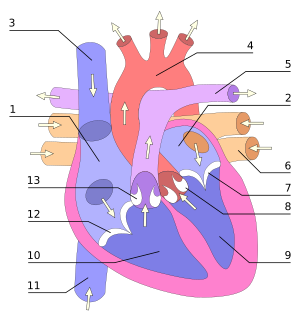
Right atrium
Left atrium
Vena cava
Aorta
Pulmonary artery
Pulmonary vein
Mitral valve (AV)
Aortic Valve
Left ventricle
Right ventricle
Vena cava
Tricuspid valve (AV)
Sigmoid valve (pulmonary)
Depleted blood oxygen by passing through the body enters the right atrium by three veins, the superior vena cava (superior vena cava), the inferior vena cava (inferior vena cava) and the coronary sinus. The blood then passes into the right ventricle. The latter pump to the lungs through the pulmonary artery (arteria pulmonalis).
After losing its carbon dioxide to the lungs and it be provided with oxygen and blood through the pulmonary veins (venae pulmonales) into the left atrium. From there the oxygenated blood enters the left ventricle. This is the main pumping chamber, designed to send blood through the aorta (aorta) to all parts of the body except the lungs.
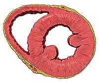
Transverse section through the ventricles (the left ventricle to the right of the image)
The left ventricle is much more massive than the right because he must exert considerable force to force the blood through the body pressure against the body, while the right ventricle serves only the lungs.
Although the ventricles are at the bottom of the atria, the two vessels through which blood leaves the heart (the pulmonary artery and aorta) at the top of the heart.
The wall is composed of heart muscle that does not tire. It consists of three distinct layers. The first is the pericardium (pericardium) that consists of a layer of epithelial cells and connective tissue. The second is the thick myocardium (myocardium) or cardiac muscle. Inside is the endocardial (endocardium), an additional layer of epithelial cells and connective tissue.
The heart needs a large amount of blood donated by the coronary arteries (whose circulation is called diastolic) left and right (arteriae coronariae), branches of the aorta.
The Heart Revolution
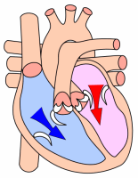
Diastole and
Atrial systole
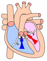
Ventricular systole
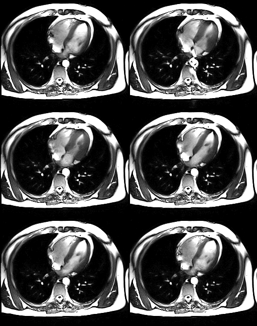
Heartbeat filmed in MRI, and only the ventricles are visible
The resting heart rate is 55 to 80 beats per minute, for a throughput of 4.5 to 5 liters of blood per minute. In total, the heart can beat more than 2 billion times in a lifetime. Everyone's beating leads a sequence of events collectively called the cardiac revolution. It consists of three major steps: the systolic atrial, ventricular systole and diastole:
During atrial systole, the atria contract and eject blood into the ventricles (fill active). Once the blood expelled from the atria, the atrio-ventricular valves between the atria and ventricles close. This prevents reflux of blood into the atria. The closure of these valves produces the familiar sound of the beating heart.
Ventricular systole involves contraction of the ventricles, expelling the blood into the circulatory system. Once the blood expelled two sigmoid valve - the pulmonary valve to the right and left aortic valve - close. Thus the blood does not flow back into the ventricles. The closure of semilunar valves produced a second heart sound sharper than the first. During the systole the atria now released, fill with blood.
Finally, the diastole is the relaxation of all parts of the heart, allowing the filling (liabilities) of the ventricles by the atria and right and left from the vena cava and pulmonary.
The heart goes 1 / 3 of the time in systole and 2 / 3 in diastole.
The rhythmic expulsion of blood and causes the pulse that can feel.
Regulation of cardiac contractions
Automation heart
The heart muscle is 'myogenic'. This means that, unlike muscle, skeletal, which requires a conscious or reflex stimulus, excites the heart muscle itself. The rhythmic contractions occur spontaneously, although their frequency may be affected by nervous or hormonal influences such as exercise or the perception of danger.
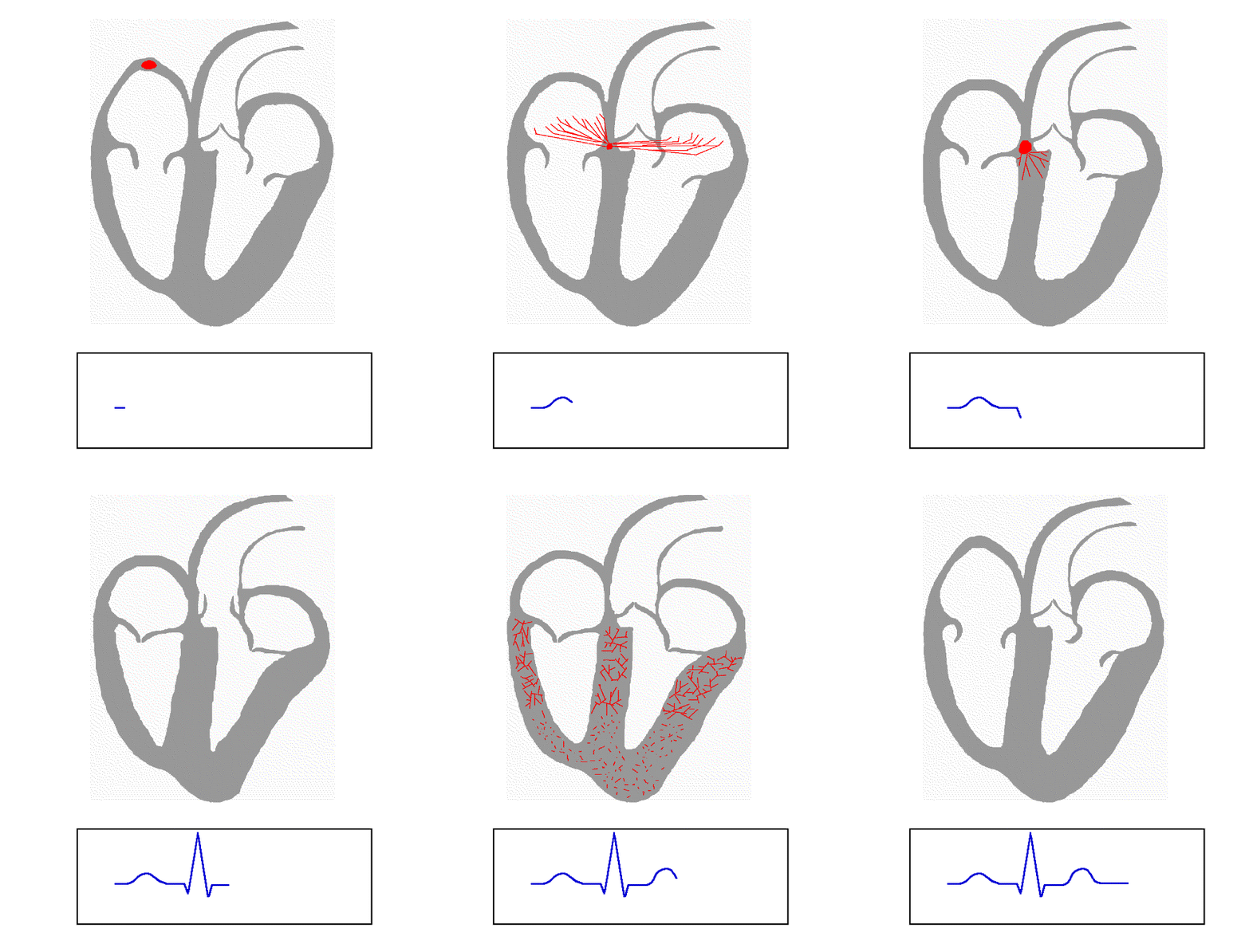
The depolarization of heart cells during a cycle
The rhythmic sequence of contractions is coordinated by a depolarization (reversing the electric polarity of the membrane by active passage of ions through it) of the sinus node or node of Keith and Flack (sinuatrialis nodes) located in the upper wall of the right atrium. The electric current induced in the order of millivolts, is transmitted throughout the atria and passes into the ventricles through the atrio-ventricular node. It spreads through the septum by the His bundle, consisting of specialized fibers called Purkinje fibers and serve as filters in case of too rapid activity of the atria. Purkinje fibers are specialized muscle fibers allowing good electrical conduction, which ensures the simultaneous contraction of the ventricular walls. The electrical system explains the regularity of the heartbeat and coordinates the atrio-ventricular contractions. It is this electrical activity is analyzed by electrodes placed on the surface of the skin and is the electrocardiogram or ECG.
Control by the central nervous system
The power and frequency of contractions are modulated by centers in the medulla oblongata, through nerves moderator cardiovascular and cardio-stimulator. These nerve centers are sensitive to blood conditions: pH, concentration of oxygen.
Hormonal Regulation
Hormones such as adrenaline and noradrenaline (hormones and the adrenergic system [Ortho] sympathetic) or thyroid hormone (T3) promotes contractility. In contrast, hormones such as acetylcholine (hormone parasympathetic or cholinergic system) slows the heartbeat.
The sympathetic nervous system in addition to its direct action on the heart in particular will cause dilation of coronary arteries (and bronchioles) that supply the heart while allowing increased blood flow and thus an increase in muscular effort can therefore increased frequency of contractions. The parasympathetic system instead will produce a constriction of coronary arteries (and bronchioles) then causes a decrease in blood flow decreased muscle power potential, acting like an "engine brake".
Diseases and treatments
- The study of heart disease called the Heart. The primary cardiac disease include:
- The CHD is coronary artery disease deprives the heart muscle of oxygen. Reversible, it can cause chest pain called severe angina pectoris (angina pectoris). The occlusion of an artery acute causes cell death of heart muscle (myocardial infarction).
- The heart failure is the progressive loss of the heart's ability to provide blood flow. It is manifested by dyspnoea (breathlessness), by edema of the legs and can go up to the acute pulmonary edema.
- the valvular heart disease: achieving valves sometimes manifested by a "heart murmur".
- The endocarditis and myocarditis are inflammations of the heart due to bacterial or viral.
- The arrhythmia of the heart is an irregular heart beat. A conduction disturbance causes bradycardia (heart or too slow).
- The pulmonary embolism is the obstruction of a pulmonary artery by a clot.
- The congenital heart diseases, that is to say, a malformation of the heart, there can be reversals of the ventricles, the atria, or both, malformation of blood vessels near the heart, or more frequently poor partitioning by the septa, particularly the non-closure of the foramen ovale between the atria.
If the coronary artery is narrowed or blocked, you can work around the place affected with coronary bypass surgery, or expand with angioplasty.
The beta-blockers are drugs that slow the beating of the heart and reduce heart needs oxygen. The nitroglycerin and other compounds that emit the nitric oxide used in the treatment of heart disease because they cause dilation of coronary vessels.
The first heart transplant was performed at Groote Schuur Hospital in Cape Town (South Africa) on 3 December 1967. Washkansky Lewis, 53, received a heart of a young woman died in a road accident. He died 18 days later from pneumonia. The surgical team was headed by Christiaan Barnard. In France, Emmanuel Vitria lived from 1968 to 1987 with a heart transplant.