Structure of DNA
DNA is a molecule duplex, ie consists of two chains arranged in antiparallel manner and with the nitrogenous bases facing each other. In three-dimensional structure, there are different levels:
Primary structure:
Sequence of nucleotide chains. It is in these channels where the genetic information, and because the skeleton is the same for all the difference in the information lies in the different sequence of nitrogenous bases. This sequence has a code, which determines an information or otherwise, as the order of the bases.
Secondary structure:
It is a double helix structure. Can explain the storage of genetic information and the mechanism of DNA replication. It was postulated by Watson and Crick, based on X-ray diffraction that Franklin and Wilkins had been made, and the equivalence of bases Chargaff, whereby the sum of adenines more guanines is equal to the sum of thymines more cytokines.
It is a double strand, right-handed or left-handed, depending on the DNA. Both chains are complementary, as adenine and guanine in a chain are joined, respectively, thymine and cytosine on the other. Both chains are antiparallel, then the 3 'end of one faces the 5' end of the counterpart.
There are three models of DNA. The DNA of type B is the most abundant and is discovered by Watson and Crick.
Tertiary structure:
Refers to how DNA is stored in a confined space to form the chromosomes. Varies depending on whether the organisms prokaryotes and eukaryotes:
In prokaryotes the DNA is folded like a super-helix, usually in circular shape and associated with a small amount of protein. The same happens in cellular organelles such as mitochondria and the chloroplasts.
In eukaryotes, since the amount of DNA from each chromosome is very large, the packing must be more complex and compact, this requires the presence of proteins such as histones and other proteins of non-histone nature (in the sperm of these Proteins are the protamines).
Double helix structure

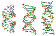
From left to right, the DNA structures A, B and Z.
DNA exists in many conformations. However, in living organisms have only been observed conformations A-DNA, DNA-B and Z-DNA. The conformation DNA adopts depends on its sequence, the amount and direction of supercoiling that show the presence of chemical modifications on the bases and conditions of the solution, such as the concentration of ions of metals and polyamines. In the three conformations, the form "B" is the most common conditions in the cells. The two DNA double helices alternatives differ in their geometry and dimensions.
The form "A" is a spiral that rotates clockwise, wider than the "B" with a minor groove surface and wider, and a slot closer and more profound. The form "A" occurs in non-physiological conditions in dehydrated form of DNA, whereas in the cell can be produced in hybrid pairings of DNA-RNA strands as well as in enzyme-DNA complex.
The DNA segments in which the bases have been modified by methylation may undergo major conformational changes and take the form "Z". In this case, the strands rotate around the propeller shaft in a spiral that turns left, the opposite of a "B" more frequent. These unusual structures can be recognized by specific proteins that bind to Z-DNA and are possibly involved in the regulation of transcription.
Quadruplex structures in

Structure of a DNA quadruplex formed by repeats on the telomere. The formation of the support structure of DNA differs significantly from the typical helical structure.
At the ends of linear chromosomes are specialized regions of DNA called telomeres. The main function of these regions is to allow the cell to replicate chromosome ends using the enzyme telomerase, since other enzymes that replicate DNA can not copy the 3 'ends of chromosomes. These specialized chromosome ends also protect DNA ends, and prevent the systems from DNA repair in the cell processes such as DNA damage that must be corrected. In human cells, telomeres are long areas of single-stranded DNA containing several thousand repetitions of a single sequence TTAGGG.
These guanine-rich sequences may stabilize chromosome ends by forming structures of stacked sets of four base units, instead of the base pairs other structures normally found in DNA. In this case, four guanine bases are flat units which are stacked one above another, to form a stable G-quadruplex structure. These structures are stabilized by forming hydrogen bonds between the ends of the bases and chelation of a metal ion in the center of each unit of four bases. can also form other structures, with the core set of four bases from either a single strand folded around the bases, or several different parallel strands so that each base contributes to the central structure.
In addition to these stacked structures, the telomeres also form large loop structures, called telomere loops or bows-T. In this case, the single DNA strands are curled in on themselves in a wide circle stabilized by proteins that bind to telomeres. At the end of the rope-T, single-stranded telomeric DNA is attached to a region double-stranded DNA because the DNA strand telomeric alters the double helix and is mated to one of the two strands. This structure is called a triple-stranded displacement loop or loop-D (D-loop).
Major and minor grooves
Animation of the structure of a section of DNA. The rules are horizontally between the two spiraling strands. Extended version

Double helix: a) Righthanded, b) Lefthanded
The double helix is a spiral right-handed, that is, each of the chains nucleotide turn right, this can be verified if you look, going upwards in the direction the segments are the threads that are in the foreground . If the two strands rotate clockwise it is said that the double helix is right-handed, and if they turn to left, left-handed (this form may appear in helices alternatives because conformational changes in DNA). But the most common conformation adopted by the DNA double helix is right-handed, turning every couple of bases on the previous approximately 36 º.
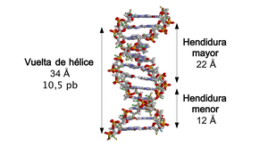
When the two DNA strands are rolled over each other (either left or right), holes or cracks are formed between a thread and the other, exposing the sides of the nitrogenous bases inside. In the most common conformation DNA adopts appear, because of the angles between the sugars of both strands of each pair of nitrogenous bases, two types of cracks around the surface of the double helix: one, the cleft or major groove, which is 22 Å (2.2 nm) wide, and the other, the groove or minor groove, which is 12 Å (1.2 nm) wide. Every turn of helix, when it has made a 360 º or what is the same from beginning to end slit slit couple minor measure by both 34th, and in each of these rounds is about 10.5 bp.

Major and minor grooves of the double helix.
The width of the slit greater implies that the ends of the bases are more accessible in this, so that the amount of exposed chemical groups is also greater which makes the differentiation between the base pairs AT, TA, CG, GC. As a result, will also be easier to accept DNA sequences from different proteins without the need to open the double helix. Thus, proteins such as transcription factors that bind to specific sequences, frequent contact with the sides of the bases exposed in the groove more. By contrast, the chemical groups that are exposed in the minor groove are similar, so that the recognition of base pairs is more difficult, for it is said that the couple split contains more information than the lower groove.
Sense and antisense
A DNA sequence is called "sense" if its sequence is the same as the sequence of a messenger RNA is translated into a protein. The sequence of the complementary DNA strand is called "antisense" (antisense). In both strands of DNA double helix can exist either sense sequences that encode mRNA as antisense, which does not encode. That is, the mRNA coding sequences are not all present on one strand, but divided between the two strands. Both in prokaryotes and in eukaryotes occurring antisense RNA sequences, but the function of these RNAs is not entirely clear. It has been suggested that antisense RNAs are involved in the regulation of gene expression by RNA-RNA pairing: the RNA antisense mRNA is complementary to aparearían, thus blocking its translation.
In a few DNA sequences in prokaryotes and eukaryotes (this is more frequent in plasmids and viruses), the distinction between sense and antisense strands is more diffuse, because they exhibit overlapping genes. In these cases, some sequences DNA has a dual function, encoding a protein when read along one strand, and a second protein when read in the opposite direction along the other strand. In bacteria, this overlap may be involved in regulating the transcription of the gene, while in viruses overlapping genes increase the amount of information that can be encoded in their tiny genomes.
Supercoiling
Structure of linear DNA molecules with fixed ends and supercoiled. For clarity, we have omitted the helical structure of DNA.
The DNA can be twisted like a rope in a process called DNA supercoiling. When DNA is in a "relaxed", a strand usually revolves around the double helix axis once every 10.4 base pairs, but if the DNA is twisted the strands may be linked more closely or more loosely. If the DNA is twisted in the direction of the helix, it is said that the positive supercoiling, and the bases are held together more closely. If the DNA is twisted in the opposite direction is called negative supercoiling, and away basis. In nature, most DNA has slight negative supercoiling that is produced by enzymes called topoisomerases. These enzymes are also required to release the torsional forces introduced in the DNA strands during processes such as transcription and replication .
Chemical modifications

cytosine 5-methyl-cytosine thymine
Structure of cytosine with and without the group methyl. After des amination, 5-methyl-cytosine has the same structure as thymine.
Database Modifications
The gene expression is influenced by the manner in which DNA is packaged into chromosomes, in a structure called chromatin. The changes of bases can be involved in DNA packaging: the regions that have a low or no gene expression usually contain high levels of methylation of the bases cytosine. For example, methylation of cytosine, producing 5-methyl-cytosine, which is important for inactivation of the X chromosome. The average level of methylation varies between organisms: the worm Caenorhabditis elegans lacking cytosine methylation, while vertebrates have a high level - up to 1% of their DNA containing 5-methyl-cytosine. Despite the importance of 5-methyl-cytosine, it can des amines to generate a thymine base. Methylated cytosines are therefore particularly sensitive to mutations. Other base modifications include the methylation of adenine in bacteria and the glycosylation of uracil to produce the "base" J "in Kinetoplastida.
DNA Damage

Benzo, the major mutagen of snuff, coupled with DNA.
DNA can be damaged by many types of mutagens, which change the DNA sequence: alkylating agents, in addition to electromagnetic radiation of high energy, like light ultraviolet and X rays. The type of DNA damage depends on the type of mutagen. For example, UV light can damage DNA dimers producing thymine, formed by cross linkage between bases pyrimidine. On the other hand, oxidants such as free radicals or hydrogen peroxide produce multiple injuries, including modifications of bases , especially guanine, and double-strand breaks (double-strand breaks). In either a human cell, about 500 bases suffer oxidative damage per day. Of these oxidative lesions, the most dangerous are double-stranded breaks, as they are difficult to repair and can produce point mutations, insertions and deletions of DNA sequence and chromosomal translocations.
Many mutagens are positioned between two adjacent base pairs, so called intercalating agents. Most molecules intercalating agents are aromatic and flat, like ethidium bromide, the daunomycin, the doxorubicin and thalidomide. For an intercalating agent can be integrated between two base pairs, they must separate, distorting the DNA strands and opening the double helix. This inhibits transcription and replication of DNA, causing toxicity and mutations. Therefore, DNA intercalating agents are often carcinogens: the benzopyrene, the acridines, the aflatoxin and ethidium bromide are well known examples. However, due to its ability to inhibit replication and DNA transcription, these toxins are also used in chemotherapy to inhibit the rapid growth of the cells cancerous.
The DNA damage initiates a response that activates different repair mechanisms that recognize specific lesions in DNA that are repaired at the time to recover the original DNA sequence. Also, DNA damage causes a shutdown in the cell cycle, which involves the alteration of numerous physiological processes, which in turn implies synthesis, transport and degradation of proteins. Alternatively, if the genomic damage is too great to be repaired, the control mechanisms induce the activation of a series of cellular pathways that culminate in cell death.