To do any exercise correctly you need to know which muscles are involved and the movements they perform.

Anatomically locating these structures allows us to identify opposing forces and how to use them to benefit our fitness routine.
When selecting exercises for the abdomen, which we include in our abdominal workout routine, know the anatomy of the major muscles that make up this region of our body.
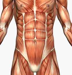
Rectus abdominis
Rectus muscle bellies pair located on either side of the midline, forming the anterior abdominal face, is covered by a robust anterior fascia that multiplies his stress. It is a very specific muscle of humans.
It originates from the top of the pubis by a short tendon of 2-3 cms. It is attached to the front of the 5th, 6th and 7th costal cartilages and xiphoid.
Are covered by a common fascia, which gives greater containment area and serves as a sheath for the displacement of the rectus abdominis muscles.
It is a muscle bellies poly gastric formed by 4 muscular tendinous bands separated by 3. The lowest is at the navel, while more than equal to the 8th rib.
Each zone receives independent nerves innervate each segment, except in the intermediate zone that is left without innervation, making a sling.
Functions of the rectus abdominis
It helps to maintain the upright position and keep the viscera in position. The contraction increases intra-abdominal pressure and helps expel the abdominal contents during bowel movements or urination. Produces flexion of the spine through the ribs. Its contraction produces unilateral lateral bending of the trunk to the same side. Your tone limits the maximum inspiration and expiration favors.

Abdominal external oblique muscle
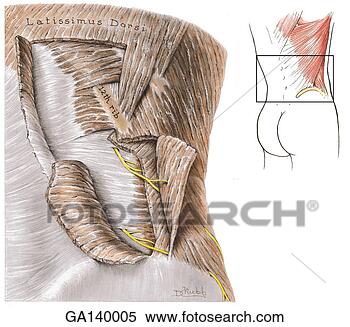
Abdomen oblique muscle is called the external oblique and superficial face and took the side of the abdomen. It is the largest of all.
It originates from the lateral aspect of ribs 5th to the 12th, through jagged scans that are interlaced with those of serratus anterior muscle and latissimus dorsi. From there the fibers pass downward and forward.
They have an extensive line of attachment, which occupies the area from the iliac crest to the outer part of the aponeurosis of the rectus abdominis. Some fibers, reaching the anterior superior iliac spine, jump up near the pubis, forming a small hole called Arc tubes, or inguinal ligament inguinal ring, through which arteries, veins, nerves and leg pass by.
Functions of the external oblique abdominal muscle
By one-sided or inclination towards the same side or rotate to the opposite side or bilaterally or trunk flexion or if the diaphragm is relaxed there is an active expiratory effort.

Many oblique muscle fibers are continuous with the oblique muscle on the other side. It works in conjunction with the oblique, so if you get the most lateral fibers of the oblique abdominal pressure occurs that contributes to the expulsion of the abdominal contents during bowel movements or urination. Abdominal internal oblique muscle
Abdomen oblique muscle is called the internal oblique and took the inner face of the external oblique muscle. It is smaller and the direction of its fibers is contrary to the oblique muscle of the same side.
It has its origin across the iliac crest and spinous processes of L5 to S1.
Its fibers are directed forwards and upwards, and are leaning progressively until the lowest fibers and earlier are transverse or horizontal.
The posterior fibers are inserted into the caudal border of the last 3 ribs on the xiphoid. The middle and lower fibers are inserting into the linea alba.
Functions of the internal oblique abdominal muscle
- By one-sided:
- or inclination towards the same side.
- or rotation to the same side.
- In bilaterally:
- or trunk flexion.
Transversus abdominis

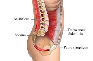
Transverse abdomens the deepest of the abdominal muscles, occupies the inner side of the abdominal wall. The fibers are horizontal.
It originates from the inside of the 5 or 6 ribs in lumbocostal ligament of vertebrae L1 to L5 in the iliac crest.
It inserts into the midline aponeurotic by a curve that is maximal at the height of the navel, which cover the posterior aspect of the rectus abdominis, being free at its lower 1 / 3. It is called Arc of Douglas.
Functions of the transversus abdominis
- Constrictor abdomen.
- Increased intraabdominal pressure.
Helps urination, defecation, vomiting, coughing, childbirth, forced expiration.